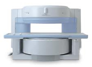
The MRI (magnetic resonance imaging) scan is the one most people will gave heard about and it is a very popular and effective way of looking at the ligaments and tendons in the shoulder.
I do use MRI scans but am moving more towards Ultra-Sound scannning because the MRI is a static scan i.e. you are just lying flat in the scanner whereas the USS allows the radiologist to move the arm around and mimic the positions that cause your pain.
MRI in conjunction with an arthrogram is very effective in looking at instability problems such as dislocations, labral tears and SLAP lesions.
In an MRI Arthrogram, a shot of a special dye is injected into the gleno-humeral joint (using the Ultra-sound machine or moving x-ray machine to make sure it goes in the right place) before you have the scan itself. The reason for putting the fluid into the joint is that it will get into any gaps, splits or tears and emphasise them whereas they could be missed on normal MRI.
 The down-side of MRI scanning is the claustrophobic effect of being inside the tube for 30 to 40 minutes. The most recent scanners are wider and the tubes are shorter so that problem has been reduced. If you really can’t face going into a normal scanner then don’t worry, we can send you to a unit where they have what’s called a ‘wide-open’ scanner where you are not enclosed.
The images from the scan are examined by the radiologist (a consultant specialising in musculo-skeletal scanning) and they write a report which gets sent to me. That process usually takes a couple of days to work through the system. When we meet for the follow-up appointment I will tell you what the scan shows and if I believe it is the cause of your symptoms and then we will decide on a course of treatment.

